Introduction to Microscopy - Part Two
Página 1 de 1
 Introduction to Microscopy - Part Two
.
.
Functions of the Major Parts of a Optical Microscope [1-3]
• Light source and Condenser: project a parallel beam of light onto the sample for illumination - Condenser gathers light and concentrates it into a cone of light that illuminates the specimen with uniform intensity over the entire view field [6].
• Sample stage with X-Y movement: sample is placed on the stage and different part of the sample can be viewed due to the X-Y movement capability
• Fine/ Focusing knobs: since the distance between objective and eyepiece is fixed, focusing is achieved by moving the sample relative to the objective lens
• Objectives: does the main part of magnification and resolves the fine details on the samples (mo ~ 10 – 100)
• Eyepiece: forms a further magnified virtual image which can be observed directly with eyes (me ~ 10)
• Beam splitter and camera: allow a permanent record of the real image from the objective be made on film (for modern research microscope).
Magnification
• To visualize any tiny object 0.1mm there is limit of unaided Human eye.
• To see micro organism much smaller than 0.1mm a system required convex lenses.
• When parallel rays pass through convex lens get converged at point called as Focal length (f) of the lens.
• Hence, a lens with shorter focal length will have higher magnification power.
• In compound microscope it will be i.e 10X, f=16mm; 40X, f=4 mm; 100X, f= 1.8mm.
• Image produced by objective lens falls on the eyepiece lens serve as object.
• Image formed in the eyepiece is perceived by one eye. The overall magnification is given as the product of the lenses and the distance over which the image is projected: M = (D *M1 * M2)/ 250 mm – (1) Where, D = projection (tube) length (usually = 250 mm); M1, M2 = magnification of objective and ocular. 250 mm = minimum distance of distinct vision for 20/20 eyes.
Resolution
• The ability to see two close objects as two distinct objects called resolution.
• The limit of resolution is the closest distance between two points at which the points still can be distinguished as separate entities.
• Magnification should be coupled with good resolution to visualize small micro organisms, else magnification alone will produce an inconclusive or blurred image.
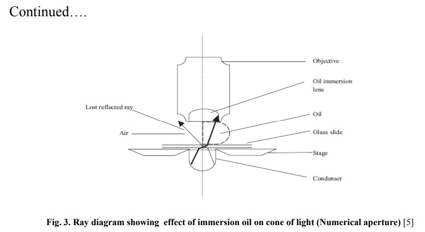
• The resolution (Fig. 2.a-c) can be calculated as shown in Eqn. 2. Maximum resolution: R = (0.061λ)/ N.A – (2) Where, 0.61 is a geometrical term, based on the average 20-20 eye, λ = wavelength of illumination, N.A. = Numerical Aperture - The N.A. is a measure of the light gathering capabilities of an objective lens. N.A. = n sin α – (3) Where, n = index of refraction of medium, α = < subtended by the lens ra
Continued…
Continued by...
att.

 Tópicos semelhantes
» Introduction to Microscopy - Part One
» Introduction to Microscopy
» Education in Microscopy and Digital Imaging - Part 2
» Microscopy in Resolution - Resolução na Microscopia
» Microscopy Categories - Categoria dos Microscópios
» Introduction to Microscopy
» Education in Microscopy and Digital Imaging - Part 2
» Microscopy in Resolution - Resolução na Microscopia
» Microscopy Categories - Categoria dos Microscópios
Página 1 de 1
Permissões neste sub-fórum
Não podes responder a tópicos|
|
|

 Início
